“Brain tumor” is a term the majority of us are familiar with, and most of us hearing it feel similarly anxious.
A brain tumor originates as a tiny cell, multiplying out of proportion and assuming odd patterns.
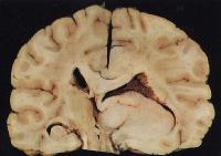
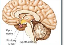
Clinically, a tumor is a type of neoplasm of the brain. Any abnormal growth of cells which forms a mass of tissue that hinders the normal functioning of the central nervous system is called a brain tumor.
An astrocytoma is a neoplasm in the brain or spinal cavity which arises out of certain glial cells present in the cerebrum of the human brain. These star-shaped cells grow to eventually form a chunk of cells referred to as a brain tumor in layperson's terms.
Astrocytoma is known to restrict its presence and growth in the human brain and spinal cord and is only very rarely known to reach to other body parts.
Since it develops out of glial cells, it’s also called a glioma in medical terminology.
An astrocyma or a glioma can be classified into two broad categories:
- Tumors with contracted areas of permeation These tumors are invasive in nature, which means they progressively spread to other sections of the human brain or the spinal column. Examples are pilocytic astrocytoma, pleomorphic xanthoastrocytoma and subependymal giant cell astrocytoma. Characteristically, these have a clearly visible outline in the CT Scans, MRIs or MRTs.

Low-grade Astrocytoma
- Tumors with scattered zones of penetration These usually develop in one of the cerebral regions. However, they are known to initiate anywhere in the Central Nervous System. For instance, low-grade astrocytoma, anaplastic astrocytoma and glioblastoma multiforme usually occur in adults, and their fundamental nature is to progressively worsen in intensity and to spread.
There is no particular age when an astrocytoma may develop. Children and adolescents are more prone to the low-grade type, whereas adults are reported to be prone to the higher grades of the disease.
Anaplastic Astrocytoma
Grading
There are multiple grading systems available to classify the tumors affecting the CNS. However, the grading system developed and employed by the World Health Organization is more widely used than most other methods. WHO’s histological guidelines categorize the grades of astrocytoma into four levels.
Grade-I is assigned to the least destructive, and subsequent grades progress with the level of aggression.
The ranking system employed by WHO is devised by considering the emergence of certain typical characteristics.
- Atypia – The level of abnormality of the cell in question.
- Mitosis – The intricacies of the cell division process by which chromosomes of a cell are divided into two identical sets.
- Endothelial Proliferation – The intensity of penetration of the neoplasm into the endothelium layer is a decisive factor in deciding the grade of an Astrocytoma. Endothelium consists of a very thin coating of cells lining the inner surface of nerves, veins and lymphatic vessels.
- Necrosis – The untimely death of cells in a living tissue due to unnatural reasons.
These features are significant in measuring the malignancy of an astrocytoma with regard to its invasion and developmental pace. Any tumor with none of these features present is Grade-I. A tumor with only one of these features is Grade-II. An astrocytoma with two of these aspects prominent is Grade-III, and one with either three or all four aspects present is Grade-IV.
Grade I and II astrocytomas are low-grade, which make up, respectively, a mere 2% and 8% of all reported astrocytomas according to research reports at WHO. Grade-III anaplastic astrocytoma accounts for 20%.
Grade-IV is the most rampant. GBM — glioblastoma multiforme — constitutes 70% of all astrocytomas reported.
As well, GBM is the most common form of cancer of the CNS and the next most common brain tumor, second only to brain metastasis. Astrocytoma is a less prevalent form of cancer in comparison to other cancers occurring in humans. However, the mortality rate is considerably high, owing to late detection and the dispersed spread of the grade III & IV astrocytomas.
Grade II Astrocytoma
Pathophysiology
Astrocytoma causes
- Undue pressure, invasion, and damage of the normal functioning of the brain.
- Lack of adequate oxygen supply to arteries and veins of the brain, termed as hypoxia.
- Competition for nutrients between the tumor and healthy brain tissues.
- Increased pressure within the skull, abnormally increasing the volume of blood and CSF (Cerebro-Spinal Fluid).
Symptoms
Symptoms vary on an individual basis depending on the severity of the astrocytoma and may comprise one or many of the following:
- Recurrent headaches
- Vision problems
- Nausea and vomiting for no apparent reason
- Decline in appetite
- Mood swings and unexplained changes in personality traits
- Diminished abilities to comprehend and learn
- Sudden occurrence of seizures or convulsions
- Difficulty in speaking
Diagnosis
A characterization of the spread, position and progressiveness can be analyzed with diagnostic techniques like a Computed Tomography Scan (CT scan) or a Magnetic Resonance Imaging (MRI) scan.
A diagnostic image will show the presence and growth of an astrocytoma and the resultant distortion of the brain or the spinal cord. A histological investigation is indispensable for determining the grade of the brain tumor.
Microscopic View of Glioblastoma Multiforme
As a preliminary diagnostic step, the physician will record the symptoms and carry out primary neurological tests, which will include tests of vision, sense of balance, coordination, and behavioral and intellectual tests.
Next, a CT scan or an MRI will be called for by the doctor. A CT scan entails taking the patient’s brain x-rays from various directions, which are then technically combined to produce a cross-sectional representation of the brain.
The response of water molecules in the patient’s brain, subjected to a varying electromagnetic field, is recorded in the form of an image in the MRI process.
In the case of a tumor being detected, a biopsy will be performed by a neurosurgeon either before or during the surgery. The pathological report of this test will enable the doctor to determine the grade and severity of the astrocytoma.
Treatment
For grade I and II astrocytomas, surgical removal normally enables the functional survival of the patient for several years. According to research data, more than 90% of patients have survived for approximately five years after a successful operation.
Low-grade astrocytoma may or may not penetrate into healthy brain tissues and are commonly known to be indolent, permitting normal neurological functions. However, they might assume a more severe form if they don’t receive prompt and necessary attention.
Even today, complete surgical elimination of grade III and IV astrocytomas has been impossible due to their tendency to scatter in various healthy tissues all over the brain and, in some cases, the spinal cord. These astrocytomas therefore have an extremely high probability of recurrence even after a successful surgical procedure.
Despite decades of therapeutic research, it is impossible to completely cure high-grade astrocytomas. A team of doctors will focus more on alleviating patients' suffering rather than on curing, and palliative care is the rational option.
There are no specific guiding principles for the prevention of this pathological condition because the specific reason for the development of astrocytomas is yet to be discovered.


